When I tell a patient that their retinal condition requires laser treatment, the reaction is almost universally one of apprehension. The concept of a laser—a high-energy beam of light—being aimed inside the delicate structure of the eye is instinctively frightening.
As a retina specialist, however, I view the laser not as a weapon, but as one of the most precise, essential, and sight-saving tools in modern medicine. It allows us to treat pathology at the microscopic level on the back wall of the eye without ever making a surgical incision.
The purpose of this article is to move past the fear of the word “laser” and explain the medical reality: the pathophysiology we are treating, the mechanism of photocoagulation, and what this therapy means for your long-term vision.
The Pathophysiology: Why We Use Laser
To understand why we need laser, we must understand how retinal disease progresses. The retina is the light-sensing tissue at the back of the eye, and it possesses the highest oxygen consumption rate of any tissue in the human body by weight. It is incredibly metabolically demanding.
In diseases like Diabetic Retinopathy or Retinal Vein Occlusion (RVO), the tiny blood vessels feeding the retina become damaged or blocked. This leads to two disastrous outcomes:
- Ischemia (Oxygen Starvation): Parts of the retina begin to suffocate. In a desperate attempt to get oxygen, the ischemic retina releases a protein called VEGF (Vascular Endothelial Growth Factor). VEGF signals the body to grow new blood vessels. Unfortunately, these new vessels are abnormal, fragile, and prone to massive bleeding inside the eye.
- Vascular Leakage: Damaged vessels become porous, leaking fluid and proteins into the retina, causing swelling (edema) that destroys vision.
The Mechanism: Understanding Photocoagulation
Retinal laser therapy is technically known as photocoagulation.
The laser is not a “cutting” tool in this context. Instead, it delivers highly focused thermal energy to specific spots on the retina. When the laser beam hits the pigmented tissue of the retina, that light energy is converted into heat. This creates a microscopic burn, or coagulation spot.
We use this thermal effect in two distinct ways:
1. Barricade and Seal (Focal Laser)
For conditions like a retinal tear or localized leakage in diabetic macular edema, we use the laser to create a barrier.
- Retinal Tears: We “spot weld” around the tear. As these laser spots heal, they form a ring of scar tissue that “glues” the retina down, preventing fluid from getting underneath and causing a full retinal detachment.
- Leaking Vessels: We target specific microaneurysms (ballooning vessels) to cauterize and seal them, stopping the leakage of fluid.
2. Reducing Oxygen Demand (Panretinal Photocoagulation – PRP)
This is used for severe proliferative diabetic retinopathy or vein occlusions where new, dangerous blood vessels are growing unchecked.
The concept here is one of necessary sacrifice. We cannot save the entire retina when blood flow is severely compromised. If we try to save everything, the entire eye may be lost to hemorrhage or detachment.
With PRP, we apply thousands of laser spots to the peripheral (side) retina—the areas that are most oxygen-starved. By sacrificing this peripheral tissue, we reduce the overall oxygen demand of the eye and stop the production of the VEGF distress signal. This causes the dangerous new vessels to regress and dry up, saving the crucial central vision.
The Procedure: A Clinical Walkthrough
Laser surgery is an in-office procedure, not an operating room event. It typically takes 15 to 30 minutes.
- Preparation: Your eye will be maximally dilated. We then apply strong anesthetic drops to completely numb the surface of the eye.
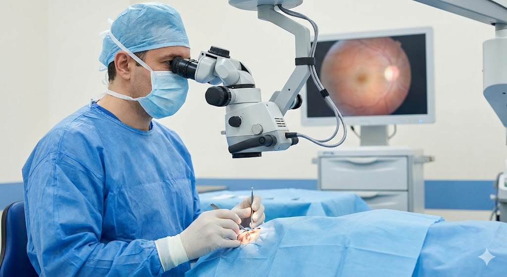
- The Contact Lens: I place a specialized gel and a contact lens on your eye. This lens keeps your eye still, keeps the eyelid open, and serves as a microscope to focus the laser beam precisely onto the retina.
- The Experience: You sit at a slit-lamp microscope. You will see very bright flashes of light—green, yellow, or red, depending on the laser used. You will hear a clicking sound with each application.
- Sensation: For focal laser, patients rarely feel anything. For extensive PRP, you may feel a “zinging” sensation, a dull ache, or pressure behind the eye as the procedure continues. It is generally well-tolerated without sedation.
Recovery and Aftercare
Immediately after the procedure, your vision will be very dark and blurry for a few hours due to the intensity of the light. This is normal “flash blindness,” similar to having a photo taken with a bright flash, but longer-lasting.
You may have a mild “eye headache” for 24 hours, easily managed with over-the-counter pain relievers. There are usually no patches required, and you can resume most normal activities the next day.
The Crucial Reality Check: Managing Expectations
As a doctor, this is the most important message I convey regarding retinal laser: Laser is usually a stabilization tool, not a restoration tool.
Patients often hope that laser surgery will return their vision to 20/20. In most cases of chronic disease like diabetes, the goal of laser is to stop the progression of the disease and prevent blindness. While some patients see improvement as swelling resolves, the primary metric of success is that vision stops getting worse.
Retinal laser is a preemptive strike against catastrophic vision loss. It is a calculated, scientifically proven intervention designed to maintain your sight for the long term.