For decades, surgery on the retina—the delicate, light-sensing tissue at the back of the eye—was considered one of the most invasive procedures in ophthalmology. It required large incisions, significant recovery time, and considerable post-operative discomfort.
Today, that narrative has changed entirely with the advent of Micro-Incision Vitrectomy Surgery (MIVS), commonly known as Sutureless Vitrectomy.
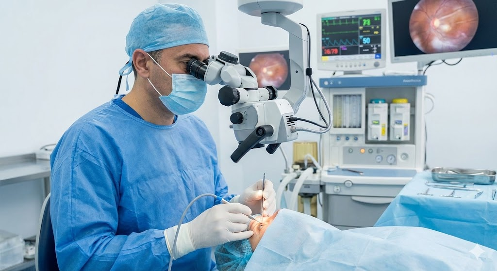
This procedure represents a paradigm shift in vitreoretinal surgery, moving from “open” surgery to a keyhole approach that prioritizes physiological stability and rapid visual recovery. Here is the medical breakdown of how we repair the retina without a single stitch.
The Anatomy: The Vitreous Chamber
To understand the surgery, you must understand the geography of the eye. The front of the eye contains the lens (treated in cataract surgery). The back roughly 80% of the eye’s volume is filled with a clear, egg-white-like gel called the vitreous humor.
When disease strikes the back of the eye—whether it is a retinal detachment, diabetic bleeding, or a macular hole—we often need to remove this gel to access the retina. This removal is called a vitrectomy.
The Evolution: The Gauge System
In medicine, needle thickness is measured in “gauge” (G). The higher the number, the thinner the instrument.
- Traditional Surgery (20-Gauge): Required incisions roughly $0.9mm$ wide. These were large enough to require sutures to close the sclera (white of the eye) and conjunctiva.
- Modern Sutureless Surgery (23G, 25G, 27G): We now use instruments as small as $0.4mm$ in diameter. These ports are smaller than the needle used for a standard blood test.
The Procedure: How MIVS Works
Sutureless vitrectomy relies on distinct bio-engineering principles that differentiate it from traditional surgery.
1. Trocar-Cannula Entry
Instead of cutting the eye open with a blade, we insert three microscopic tubes called trocars through the sclera. These serve as “ports” or tunnels.
- The Biophysics: We insert these trocars at an angle. This creates a “beveled” or tunneled wound architecture. When the instruments are removed, the internal pressure of the eye presses the tunnel roof against the floor, effectively self-sealing the wound like a valve.
2. High-Speed Vitreous Cutters
Once inside, we use a vitreous cutter that oscillates at incredibly high speeds (up to $10,000$ cuts per minute). This allows us to shave away the vitreous gel safely without pulling on the retina (traction), which is crucial for preventing iatrogenic tears.
3. Membrane Peeling and Laser
With the gel removed, we can perform delicate maneuvers:
- Peeling: Removing microscopic membranes (like epiretinal membranes) from the macula using forceps.
- Endolaser: Applying laser burns to seal tears or treat diabetic vessels.
4. Tamponade (The “Internal Cast”)
The eye cannot remain empty. At the end of surgery, we often fill the vitreous cavity with a tamponade agent to hold the retina in place while it heals:
- Sterile Air or Gas: This is absorbed by the body over weeks.
- Silicone Oil: Used for complex detachments; requires a second minor procedure to remove later.
Clinical Indications: When is this used?
Sutureless vitrectomy is the gold standard for several sight-threatening conditions:
- Retinal Detachment: Reattaching the sensory retina to the back wall of the eye.
- Diabetic Retinopathy: Clearing blood (vitreous hemorrhage) and removing scar tissue caused by diabetes.
- Macular Holes: Repairing a break in the very center of your vision.
- Floaters: In severe cases, removing the opacities within the vitreous gel.
The Patient Advantage: Why “Sutureless” Matters
The elimination of sutures is not just about convenience; it changes the physiology of recovery.
- Reduced Astigmatism: Sutures tighten the eye tissue, warping the cornea and causing astigmatism. Sutureless ports leave the corneal shape largely unchanged.
- The “Quiet” Eye: There is significantly less inflammation and redness. Patients often experience a “white eye” within days rather than weeks.
- Comfort: No sensation of foreign bodies (stitches) rubbing against the eyelid.
- Efficiency: Reduced surgical time means less trauma to the eye tissues.
Post-Operative Reality
While the surgery is minimally invasive, the recovery often requires patient cooperation, specifically regarding head positioning.
If a gas bubble is used, you may need to maintain a specific head posture (e.g., face down) for a few days. This allows the bubble to float up and press against the specific area of the retina that needs to heal.
Conclusion
Sutureless vitrectomy is a triumph of microsurgical engineering. It allows us to perform major reconstruction on the most sensitive neural tissue in the human body through openings smaller than a grain of sand.
If you have been diagnosed with a retinal condition, understand that the technology used to treat you today is safer, faster, and more precise than ever before.